How do ear infections happen?
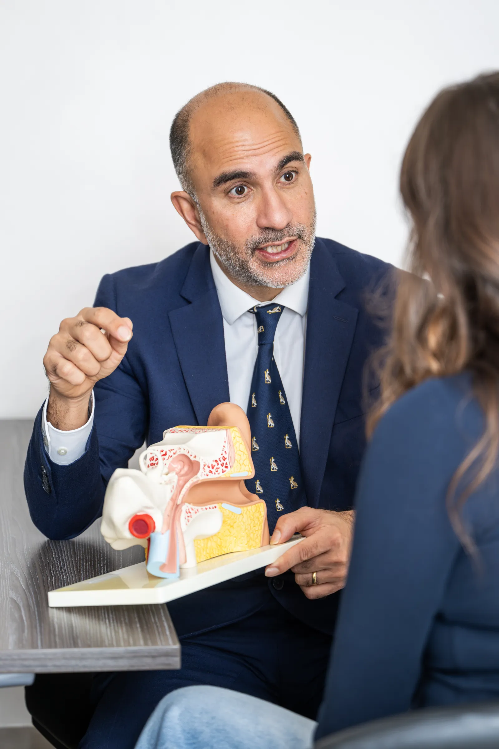
There are three anatomical parts of the ear which are connected together:
- The outer ear — comprising the ear canal down to the eardrum.
- The middle ear beyond the eardrum — a box-like structure containing the three bones of hearing (the hammer, the anvil and the stirrup) which connects with the back of the nose via the Eustachian tube.
- The inner ear — containing the balance and hearing organs.
All three can get infections, either separately or together, and this will lead to different symptoms and signs depending on which part of the ear is affected. The treatment of the infections is therefore different too, depending on which part(s) of the ear are involved.
Infection of the outer ear is called otitis externa. Infection of the middle ear is called otitis media. Infection of the inner ear is called labyrinthitis.
Otitis externa
This is the most common type of ear infection, and is basically a superficial skin infection in the blind-ending tunnel of the external ear canal.
It is more commonly seen in people with pre-existing skin problems such as eczema and psoriasis, where the skin barrier to infection is weakened, but also in people who swim, particularly in polluted water. That is why it is often called ‘swimmer’s ear’.
Other factors that can make this infection more common are attempting to clean the ears with fingers, or worse still with sharp implements such as hairgrips, paper clips or particularly cotton buds. These often damage and tear the delicate ear canal skin and allow infection to take hold once the barrier effect of the skin to bacteria is lost.
Do not put anything smaller than your elbow in your ear (think about it…!)
In everybody, the skin of the ear canal very slowly moves out of the ear over time, and this allows it to push wax out of its own accord. This clever piece of anatomy means that most people’s ears are self-cleaning, and don’t therefore need any sharp instruments stuck into them to help the wax out.
Symptoms
The commonest symptom is pain, particularly if the ear is touched or pulled around. The infection may spread into the lymph glands around the ear and the tissues around the jaw and face, so chewing may be painful too.
The ear often weeps or discharges and may itch. As the ear canal swells and becomes blocked with the discharge, you may feel deaf in the affected ear too.
Treatment
It is important to get an accurate diagnosis of where the infection in your ear is coming from before commencing treatment. Your GP may be happy to do this, but if there is a lot of swelling and discharge it may be very difficult to accurately diagnose the cause without the specialist equipment and expertise of an ENT Surgeon.
Once accurately diagnosed, the main treatment for otitis externa is eardrops or sprays. If the infection is mild or caught very early, these can be just a drying agent such as EarCalm spray, but usually will involve antibiotic and steroid combination eardrops such as Sofradex and Otomize. If the ear is very swollen and full of discharge, careful cleaning and removal of the discharge with a microscope and tiny vacuum cleaner will be required. This needs specialist ENT input, and may be necessary several times over the first two or three weeks before full recovery occurs.
During treatment, and in the next six weeks or so as the skin of the outer ear regenerates, it is very important to keep the ear scrupulously dry — either with earplugs or homemade earplugs of clean cotton wool smeared with Vaseline jelly — every time you bath or shower. You should avoid swimming in this period too.
Occasionally, if the infection doesn’t respond to the eardrops or spreads, oral antibiotics are prescribed. If the whole ear becomes swollen (perichondritis) and/or you become unwell as a result of spreading infection, admission to hospital for intravenous antibiotics may be required.
Very occasionally, this infection can spread to involve the bone of the ear canal and surrounding skull, causing osteomyelitis (‘necrotising otitis externa’). This is very uncommon in normally healthy people, but should be excluded in those with underlying immune problems (such as diabetes) or those on drugs that suppress the immune system. It requires careful specialist treatment and regular review until it settles, often after a prolonged period.
Otitis media
As with otitis externa, it is important to get an accurate diagnosis of where the infection is coming from before commencing treatment. Your GP may be happy to do this, but if there is a lot of swelling and discharge it may be very difficult to diagnose accurately without the specialist equipment and expertise of an ENT Surgeon.
(Note: the features described here are those of acute otitis media. If the infection persists beyond two weeks it is correctly termed chronic suppurative otitis media, which is beyond the scope of this article.)
This is an infection of the middle ear space, containing the three bones of hearing, which is connected to the back of the nose via the Eustachian tube.
Acute otitis media is more common in children, and particularly infants — more than 50% of cases are in the under-fives, and it’s more common in boys.
Infection most commonly first occurs when children first go to nursery or school, which is usually the first time their immature immune systems are exposed to lots of viruses and bacteria. This is combined with the fact that the Eustachian tube in children is straight and short, which means bugs can get from the nose to the middle ear in double-quick time.
Factors that may help prevent children getting acute otitis media include: breastfeeding for the first six months, avoidance of dummies, giving children pneumococcal and influenza vaccines, treating acid reflux, and particularly avoidance of parental smoke.
Symptoms
The commonest symptom is pain in and around the ear, but this time usually without the added pain on manipulation or pulling of the ear. The child may be noticeably hard of hearing due to the build-up of infected mucous in the middle ear.
Often there is also a recent history of a cough or cold with a runny nose, and the child may have a raised temperature and be irritable and not sleeping. They may hit that side of the head on their cot, or pull at the ear.
The ear may not discharge at first, as the infection is bottled up in the middle ear and confined by the intact eardrum. If it doesn’t clear over the first few days, the pressure in the middle ear may slowly rise (and with it the pain), and the eardrum often eventually bursts, releasing a rush of pus. With this release of pressure the child is often much improved, in less pain and less irritable, and the temperature starts to reduce as the infection settles.
Once the infection is under control the eardrum usually heals, but this cycle of infection and perforation may recur several times.
Very occasionally, the infection may spread into the air cells in the skull behind the ear, in the mastoid region (causing mastoiditis). This causes pain and swelling behind the ear, which may push the ear away from the side of the head. Even more unusually, the infection may spread into the skull and brain, causing meningitis and a brain abscess, but these are very rare. If these complications are suspected, hospital admission is essential.
Treatment
As it is so common, there has been a lot of research into the best treatment of acute otitis media, particularly by GP colleagues around the world.
Figures from large studies show that 50% of all cases of ear pain in children resolve without any treatment within three days, and 90% resolve within seven days. It is therefore important not to over-treat this condition with antibiotics, particularly given the mounting problems worldwide with antibiotic resistance.
A reasonable plan is to treat children initially (for 48 to 72 hours) with simple painkillers such as paracetamol and ibuprofen, and if on review they are no better or are getting worse, then to consider antibiotics at that stage.
If the pain and pressure get worse and the eardrum doesn’t burst spontaneously, occasionally a short general anaesthetic is required to make a small nick in the eardrum to let the infected mucous out (myringotomy). Often at the same time a small, short-term drainage tube (a grommet) is inserted to allow the mucous to drain for a short period.
Long-term outlook
Acute otitis media may be recurrent in children, until the immune system becomes more able to fight infection and the anatomy of the ear and Eustachian tube matures. Once the child is past around eight years old, the problems have usually started to resolve.
Sometimes after the infection has settled (and the eardrum healed if it had previously perforated) the child is left with a collection of sterile mucous in the middle ear — so-called ‘glue ear’ (see our Glue Ear and Grommets leaflet). In this situation the child is otherwise well, but often has issues with hearing, attention and balance due to the muffling effect of the fluid. This stagnant fluid also makes a further episode of acute otitis media more likely, as it is easily infected next time the child has a cough or cold.
Grommets are often a good idea in this scenario if the middle ear fluid is persistent and doesn’t settle after a period of ‘watchful waiting’ supervised by your GP or us (i.e. for more than three months).
If the infections are multiple (three or more episodes in six months, or four or more in a year) but the ear returns to normal between episodes, it may also be an option to insert a grommet to allow the middle ear to drain all the time. This is not an ideal option, however, as a grommet means there is a constant connection between the middle ear and the outside world, with the barrier effect of the eardrum lost. Grommets for this particular condition can therefore make things worse, so careful consideration and discussion is necessary in these cases.
Labyrinthitis
This is inflammation in the inner ear (cochlea) and/or balance organ (utricle, saccule and semicircular canals), which are intimately connected to each other inside the skull adjacent to your outer ear.
The cause is often not certain, but it is thought to be a viral infection reaching the inner ear through your blood after a common cold or flu. There is often a history of such an illness before the symptoms come on. It may also be due to bacterial infection as part of a generalised brain infection or meningitis, may occur after a head injury, or as a side effect of medication (check the drug information leaflet for recognised side effects if you are unsure).
This infection usually clears itself without any curative medication. There is no evidence that antibiotics or antiviral medication make any difference. Symptoms usually largely settle within two weeks, but it can take up to six weeks to feel back to normal. Even then, you may still feel off balance for up to 12 to 18 months after an episode. Overall recovery depends on the degree of rehabilitation entered into (see Treatment below), as well as your age and general fitness and balance function before the infection struck.
Symptoms
The main symptom is severe dizziness due to the effect on the balance organ structures, but depending on the degree of infection of the inner ear, you may also experience hearing loss and tinnitus.
If the symptoms are purely dizziness, you may hear this condition described as vestibular neuronitis. This implies that the balance organ and nerve running from the inner ear to the brain are predominantly affected and the inner ear has been spared, but it is essentially the same as labyrinthitis.
The dizziness is usually rotatory — the feeling that you are moving and spinning when you are not, akin to coming off a fast merry-go-round. You or your loved ones may notice your eyes flickering (called nystagmus) as you try to focus during the worst hours of the infection, a result of the disturbed messages being sent from your brain to your eyes.
You will often feel and be sick, and sometimes these symptoms can be so bad that it is difficult to lift your head off the pillow. If the symptoms are this severe, a short admission to hospital may be necessary to give you strong anti-sickness drugs and fluids via injection or into a vein to prevent dehydration while your body recovers.
Treatment
The main priority once you start to experience these symptoms is to seek medical help at the earliest opportunity. In the age group that predominantly gets this infection (30 to 60 years, peaking between 40 and 50) there is a risk of stroke, which needs to be excluded by your GP or via an Accident & Emergency assessment.
Once the diagnosis is made, the main treatment is supportive — to help relieve the dizziness and nausea via anti-sickness medication. This is often the same as the drugs used for travel sickness, such as cinnarizine (Stugeron) and prochlorperazine (Stemetil or Buccastem). These can be taken as tablets, but Buccastem dissolves next to your gum under the lip so doesn’t need to be swallowed. If you are unable to keep anything down, these drugs can be given by injection into a muscle or vein.
Once the worst of the dizziness settles, it is important to establish a course of vestibular rehabilitation. The most common form suggested is Cawthorne-Cooksey exercises. Rehabilitation is often supervised by specialised balance therapists or physiotherapists, to whom we can refer you. These exercises challenge your balance system in an increasingly forceful way to encourage recovery of the damaged part of the inner ear, and to establish compensation from other parts of the balance system in your brain, eyes, muscles and joints.
The general advice while recovering is to be as active as you can (without risking falling over or hurting yourself) in order to encourage compensation in the same way as the rehabilitation exercises do. This may require modifying your home environment (avoiding stairs, fitting props and handrails as reassurance) and the use of a stick or frame while out walking during recovery.
A positive attitude and ‘can do’ ethos is important to encourage recovery. Sitting immobile and expecting the condition to right itself without hard work on your part is not helpful — and there are colleagues of ours, trained in this aspect of treatment, who can help you.