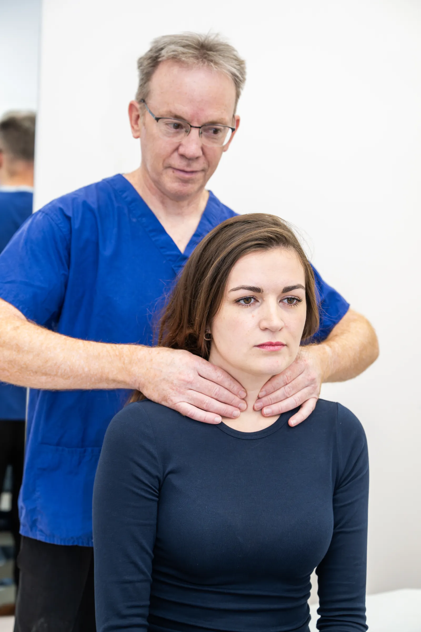
The thyroid gland sits in the centre of the neck below the voice box, consisting of two lobes connected by an isthmus, shaped like a butterfly over the windpipe. Some individuals have a pyramidal lobe above the isthmus — an embryological remnant. If this developmental path fails to close, it can form a thyroglossal duct cyst. A normal thyroid remains small and imperceptible unless enlarged.
Benign thyroid lumps and swellings
A thyroid goitre is a general enlargement of the thyroid gland, affecting one lobe or the entire gland. The most common type is a multinodular goitre, where the thyroid produces numerous small cysts or nodules causing enlargement. Causes include overactive or underactive thyroid function, puberty, pregnancy, iodine deficiency, and autoimmune conditions. These often require no treatment unless causing compressive symptoms or needing continual medical management. Sometimes a single large nodule affects only part of the thyroid. Anyone with thyroid enlargement should undergo blood tests to assess thyroid function.
Compressive symptoms of a goitre include:
- neck swelling
- difficulty breathing
- neck tightness
- swallowing problems
- a sensation of choking when lying backwards
- coughing or voice changes (rare)
Depending on the goitre’s size and symptoms, patients may receive a hemithyroidectomy (removing half the thyroid and the isthmus), an isthmectomy (removing the middle portion), or a total thyroidectomy (complete gland removal). Your surgeon will discuss the optimal option, considering symptom severity and risk factors.
Thyroid cysts are simple benign cysts within the gland, occurring singly or multiply. They may enlarge, stabilise or shrink, potentially causing compressive symptoms or noticeable neck lumps. Large cysts can be drained, but may recur, requiring surgical removal.
Thyroid nodules appear singly or multiply. Ultrasound grading and a possible needle biopsy determine their nature — non-cancerous or cancerous. Concerning ultrasound findings warrant needle testing to establish whether nodules are benign, possibly cancerous, or definitely cancerous. Nodules present as compressive symptoms or neck lumps.
Thyroid cancer
Thyroid cancer incidence is rising due to improved detection, making it the most common endocrine cancer, with an incidence rate of 9 per 100,000. Five main subtypes exist; two constitute roughly 90% of cases, with an excellent prognosis — over 95% survival twenty years after diagnosis. Thyroid cancers typically present as painless neck lumps in the gland or lymph nodes. Rarely, they cause pain, hoarseness or swallowing difficulties. Any suspected thyroid cancer requires discussion at a thyroid multidisciplinary team (MDT) meeting; our consultants Robert Hone and Ali Al-Lami participate in the East Kent thyroid and parathyroid MDT.
Papillary thyroid cancer comprises approximately 70% of thyroid cancers. Usually ‘well differentiated’, it grows slowly without aggressive behaviour, remaining stable, and the prognosis is excellent, with over 95% twenty-year survival. Occasionally, poorly differentiated variants grow faster, behaving more aggressively with a worse prognosis. When spreading occurs, it typically affects neck lymph nodes, which respond well to treatment.
Follicular thyroid cancer represents roughly 20% of thyroid cancers, with an excellent prognosis exceeding 95% at twenty years. However, this cancer spreads through the bloodstream, making distant deposits more common and harder to treat. Depending on the treatment received, thyroglobulin blood-marker monitoring follows.
Medullary thyroid cancer comprises approximately 5% of thyroid cancers, and may have a genetic predisposition. Affected patients undergo genetic screening for changes that increase susceptibility to this cancer or other tumours. Post-treatment monitoring uses calcitonin, a blood tumour marker produced by medullary thyroid cancer cells.
Lymphoma of the thyroid gland accounts for roughly 5% of thyroid cancers. It grows rapidly but responds excellently to treatment. Following diagnosis, patients transfer to a lymphoma MDT, with Haematology Department oversight for chemotherapy and/or radiotherapy.
Anaplastic thyroid cancer comprises less than 1% of thyroid cancers. Unfortunately it is very aggressive, with a poor prognosis, growing rapidly and invading nearby structures while metastasising extensively. Surgery often proves impossible, and chemotherapy and radiotherapy rarely affect this cancer. Fewer than 10% survive beyond one year after diagnosis.
Optimal thyroid cancer treatment involves surgical removal of the tumour. Depending on individual risk factors, cancer type, stage and size, additional treatment may include radioactive iodine and, rarely, radiotherapy with or without chemotherapy. Systemic chemotherapy has limited application in thyroid cancer, used only in specific instances. Regular blood tests monitoring thyroglobulin and thyroglobulin antibodies detect remaining thyroid tissue; increasing markers suggest residual or recurrent cancer, necessitating further investigation or treatment.
Investigations to determine the diagnosis
Anyone with thyroid lumps requires ultrasound examination, with or without a needle biopsy. Ultrasound determines the nature of the lump or swelling, graded per British Thyroid Association (BTA) guidelines from U1 to U5:
- U1 – benign (non-cancerous) normal thyroid gland
- U2 – benign (non-cancerous) nodule
- U3 – indeterminate nodule (around 20% chance of cancer)
- U4 – suspicious nodule (around 50% chance of cancer)
- U5 – thyroid cancer (99% being cancer)
Per BTA guidelines, nodules exceeding one centimetre graded U3, any U4, or U5 nodules require fine-needle aspiration cytology (FNAC) to examine the cells within nodules and determine cancer risk. A fine needle removes cells for microscopic assessment. Concerns about lymphoma or anaplastic thyroid cancer warrant a core needle biopsy, using larger needles for tissue removal and immunological analysis, occasionally requiring an open biopsy. FNAC results use the Thy grading:
- Thy 1 – non-diagnostic sample, possibly requiring repetition
- Thy 2 – benign
- Thy 3a – indeterminate nodule with atypical cells (around 20% chance of cancer)
- Thy 3f – indeterminate nodule with follicular cells (around 30% chance of cancer)
- Thy 4 – suspicious for thyroid cancer (roughly 60% cancer risk)
- Thy 5 – thyroid cancer (over 99% likelihood)
Thy 3 and above nodules require thyroid MDT discussion and typically surgical removal, to confirm the diagnosis and treat the nodule or cancer. The surgery offered depends on the Thy grade, size, position and appearance of the nodule. Sometimes, depending on individual factors, ultrasound surveillance (with or without repeat needle biopsy) is used to monitor nodules.
The procedure
Thyroid surgery typically requires an overnight hospital stay. Very rarely, it occurs as day surgery with same-day discharge. Occasionally, two or more hospital nights follow if low calcium levels develop and are difficult to normalise. A general anaesthetic is used. Depending on the type of thyroid swelling, the reason for surgery, or the location of the lump, procedures include:
- Hemithyroidectomy – removal of half (one side) of the thyroid
- Total thyroidectomy – complete removal of the thyroid
- Isthmectomy – removal of the middle portion of the thyroid only
Post-operative information
Patients typically have wound drains (small plastic tubes collecting excess fluid) removed the next morning. Skin sutures (possibly dissolvable) or skin clips need removal at the GP surgery after 48 hours, with steri-strips applied. A post-operative clinic follow-up occurs around 2 weeks for cancer procedures, or 4 to 6 weeks for benign cases. A repeat nasal endoscopy in clinic checks vocal cord movement. Thyroid function tests approximately 10 weeks post-operatively assess remaining gland function and adequate thyroid hormone replacement levels. Thyroid cancer patients undergo East Kent thyroid MDT discussion after the procedure, and higher thyroxine doses may be requested to suppress brain-produced thyroid-stimulating hormone.
Radioactive iodine treatment
Following thyroid cancer surgery and East Kent thyroid MDT discussion, higher-risk patients may receive radioactive iodine treatment. This involves consuming radioactive iodine, which is absorbed by remaining thyroid cells and thyroid cancer cells; the radioactive iodine then destroys these cells, ideally eliminating remaining thyroid cancer from your body. A multi-day hospital stay allows radioactive waste collection. Children should not be in contact with you for up to two weeks after treatment. There are few side effects; the treatment is very safe and effective. Post-iodine scanning assesses body uptake. One year after treatment, ultrasound and blood tests determine risk stratification into low-, intermediate- and high-risk groups, guiding further investigations and follow-up.
Risks of thyroid surgery
- Like any procedure, there are bleeding and infection risks, at around 1–2% incidence.
- Scarring — small scars at the front of the neck result. Scars are hidden within skin creases for less visibility; healed scars measure approximately 4–5 cm long, though very large thyroids may require longer incisions.
- Recurrent laryngeal nerve damage — this affects the nerve to the voice box. Damage causes potential hoarseness, at roughly 1% risk, with half being temporary. Removing the entire thyroid gland carries roughly a 1-in-10,000 chance of damage to both nerves, potentially causing immediate post-operative breathing problems — this is exceptionally rare.
- Hypothyroidism (low thyroid levels) affects up to 5% of half-thyroid removal patients, when the remaining half cannot meet the body’s demands; thyroxine supplementation becomes necessary. Complete thyroid removal requires lifetime thyroid supplements.
- Hypocalcaemia (subnormal body calcium levels) can follow complete thyroid removal. Temporary occurrence affects up to 10%, with less than 1% requiring long-term (over 3–6 months) treatment. This happens because the thyroid blood supply feeds the parathyroid glands — interruption causes temporary gland dysfunction. A minimum of two calcium checks follow a total or completion thyroidectomy.
- A hemithyroidectomy may require further surgery for:
- discovery of worrying pathology
- significant growth
- development of symptomatic compressive symptoms
- emergence of a new lump
- swallowing problems or hoarseness
If you would like to book an appointment to discuss your symptoms, please get in touch with our Practice Management Team by telephone on 01233 564455 or by email at info@kentandsussexent.com.